
vivi
with Anatomy & Physiology Lab Course s
Author Terry Martin’s forty years of teaching anatomy and physiology courses, authorship of three laboratory manuals, and
active involvement in the Human Anatomy and Physiology Society (HAPS) drove his determination to create a laboratory
manual with an innovative approach that would benet students. The Laboratory Manual for Human Anatomy & Physiology
includes a cat version, a fetal pig version, and for the rst time, a rat version. Each of these versions includes sixty-one
laboratory exercises, three supplemental labs found online, and six cat, fetal pig, or rat dissection labs. A main version with
no dissection exercises is also available. All four versions are written to work well with any anatomy and physiology text.
Preface
In Touch
Martin Lab Manual Series . . .
IN TOUCH with Anatomy & Physiology Lab Courses
▶
NEW! Available in 4 Versions: main (no
dissection), cat dissection, fetal pig
dissection, and rat dissection.
▶
Incorporates learning outcomes and
assessments to help students master
important material!
▶
NEW! Pre-Lab assignments are printed
in the lab manual. They will help
students be more prepared for lab and
save instructors time during lab.
▶
Clear, concise writing style facilitates
more thorough understanding of lab
exercises.
▶
BIOPAC
©
exercises use hardware and
software for data acquisition, analysis,
and recording.
▶
NEW! Ph.I.L.S. 4.0 included and physiology lab
simulations interspersed throughout make otherwise
difcult and expensive experiments a breeze through
digital simulations.
▶
Cadaver images from Anatomy & Physiology
Revealed
®
(APR) are incorporated throughout the
lab. Cadaver images help students make the
connection from specimen to cadaver.
▶
Micrographs incorporated throughout the lab aid
students’ visual understanding of difcult topics.
▶
Instructor’s Guide is annotated for quick and
easy use by adjuncts and is available online at
www.mhhe.com/martinseries2.
mar53064_FM.indd 6 9/14/11 8:43 AM

viivii
In Touch
with Student Needs
▶
The procedures are clear, concise, and easy to follow.
Relevant lists and summary tables present the contents
efciently. Histology micrographs and cadaver photos
are incorporated in the appropriate locations within the
associated labs.
▶
NEW! The pre-lab section now includes quiz
questions. It also directs the student to carefully
read the introductory material and the entire lab
to become familiar with its contents. If necessary, a text-
book or lecture notes might be needed to supplement the
concepts. A visit to www.mhhe.com/martinseries2 will
provide a list of animations from Anatomy & Physiol-
ogy Revealed
®
(APR) and LabCam videos to review be-
fore answering ve or more fundamental laboratory
questions for that particular lab.
▶
Terminologia Anatomica is used as the source for universal
terminology in this laboratory manual. Alternative names
are included when a term is introduced for the rst time.
▶
Laboratory assessments immediately follow each labora-
tory exercise.
▶
Histology photos are placed within the appropriate labo-
ratory exercise.
▶
A section called “Study Skills for Anatomy and Physi-
ology” is located in the front of this laboratory manual.
This section was written by students enrolled in a Human
Anatomy and Physiology course.
▶
Critical Thinking Activities are incorporated
within most of the laboratory exercises to
enhance valuable critical thinking skills that
students need throughout their lives.
▶
Cadaver images are incorporated with dissection labs.
In Touch
with Instructor Needs
▶
The instructor will nd digital assets for use in creating
customized lectures, visually enhanced tests and quizzes,
and other printed support material.
▶
A correlation guide for Anatomy & Physiology Re-
vealed
®
(APR) and the entire lab manual is located on
the lab manual’s website at www.mhhe.com/martin
series2. Cadaver images from APR are included within
many of the laboratory exercises.
▶
Some unique labs included are “Scientic Method
and Measurements,” “Chemistry of Life,” “Fetal Skel-
eton,” “Surface Anatomy,” “Diabetic Physiology,” and
“Genetics.”
▶
The annotated instructor’s guide for Laboratory Manual
for Human Anatomy and Physiology describes the pur-
pose of the laboratory manual and its special features,
provides suggestions for presenting the laboratory exer-
cises to students, instructional approaches, a suggested
time schedule, and annotated gures and assessments.
It contains a “Student Safety Contract” and a “Student
Informed Consent Form.”
▶
Each laboratory exercise can be completed during a
single laboratory session.
In Touch
with Educational Needs
▶
Learning outcomes with icons
have matching assess-
ments with icons
so students can be sure they have
accomplished the laboratory exercise content. Outcomes
and assessments include all levels of learning skills:
remember, understand, apply, analyze, evaluate, and
create.
▶
Assessment rubrics for entire laboratory assessments are
included in Appendix 2.
In Touch
with Technology
▶
Physiology Interactive Lab Simulations
(Ph.I.L.S. 4.0) is included with the lab man-
ual. Eleven lab simulations are interspersed
throughout the lab manual. The correlation
guide for all of the simulations is included
in Appendix 3.
▶
BIOPAC
©
exercises are included on
four different body systems. BIOPAC
©
systems use hardware and software for data acquisi-
tion, analysis, and recording of information for an
individual.
mar53064_FM.indd 7 9/14/11 8:43 AM

Engaging Presentation
Materials for Lecture and Lab
McGraw-Hill ConnectPlus Anatomy & Physiology is a
web-based assignment and assessment platform that gives
students the means to better connect with their coursework,
with their instructors, and with the important concepts that
they will need to know for success now and in the future.
With Connect Anatomy & Physiology, instructors can
deliver assignments, quizzes and tests easily online. Stu-
dents can practice important skills at their own pace and on
their own schedule. With Connect Anatomy & Physiology
Plus, students also get 24/7 online access to an eBook—an
online edition of the text—to aid them in successfully com-
pleting their work, wherever and whenever they choose
www.mhhe.com/martinseries2
New! All content in Connect is
correlated to HAPS Learning Outcomes.
viii
mar53064_FM.indd 8 9/14/11 8:43 AM

McGraw-Hill Higher Education and Blackboard
®
have teamed up. What does this mean for you?
1. Your life, simplied.
Now you and your students can access McGraw-
Hill’s Connect™ and Create™ right from within your Blackboard course –
all with one single sign-on. Say goodbye to the days of logging in to multiple
applications.
2. Deep integration of content and tools.
Not only do you get
single sign-on with Connect™ and Create™, you also get deep integration
of McGraw-Hill content and content engines right in Blackboard. Whether
you’re choosing a book for your course or building Connect™ assignments,
all the tools you need are right where you want them – inside of Blackboard.
3. Seamless Gradebooks.
Are you tired of keeping multiple gradebooks
and manually synchronizing grades into Blackboard? We thought so. When
a student completes an integrated Connect™ assignment, the grade for that
assignment automatically (and instantly) feeds your Blackboard grade center.
4. A solution for everyone.
Whether your institution is already using
Blackboard or you just want to try Blackboard on your own, we have a
solution for you. McGraw-Hill and Blackboard can now offer you easy access
to industry leading technology and content, whether your campus hosts it, or
we do. Be sure to ask your local McGraw-Hill representative for details.
ix
mar53064_FM.indd 9 9/14/11 8:43 AM

xx
Guided Tour Through A Lab Exercise
T
he laboratory exercises include a variety of special features that are designed to stimulate interest in the subject matter,
to involve students in the learning process, and to guide them through the planned activities. These features include the
following:
Introduc t ion The introduction describes the subject
of the exercise or the ideas that will be investigated. It
includes all of the information needed to perform the
laboratory exercise.
Materials Needed This section lists the
laboratory materials that are required to complete the
exercise and to perform the demonstrations and learning
extensions.
Pre-Lab The pre-lab includes quiz questions and directs the student to carefully read introductory material and examine
the entire laboratory contents after becoming familiar with the topics from a textbook or lecture. Students will also be
directed to visit www.mhhe.com/martinseries2 to obtain a list of correlated Anatomy and Physiology Revealed
®
animations and LabCam videos. After successfully answering the pre-lab questions, the student is prepared to become
involved in the laboratory exercise.
Procedure The procedure provides a set of
detailed instructions for accomplishing the planned
laboratory activities. Usually these instructions are
presented in outline form so that a student can proceed
efciently through the exercise in stepwise fashion.
The procedures include a wide variety of laboratory
activities and, from time to time, direct the student to
complete various tasks in the laboratory assessments.
There are also separate procedures in 11 labs that
utilize Ph.I.L.S. 4.0.
Purpose of the Exercise
The purpose provides a statement about the intent of the
exercise—that is, what will be accomplished.
Safety A list of safety guidelines is included inside
the front cover. Each lab session that requires special
safety guidelines has a safety section. Your instructor
might require some modications of these guidelines.
Learning Outco mes The learning outcomes list
what a student should be able to do after completing
the exercise. Each learning outcome will have matching
assessments indicated by the corresponding icon
in
the laboratory exercise or the laboratory assessment.
mar53064_FM.indd 10 9/14/11 8:43 AM
xixi
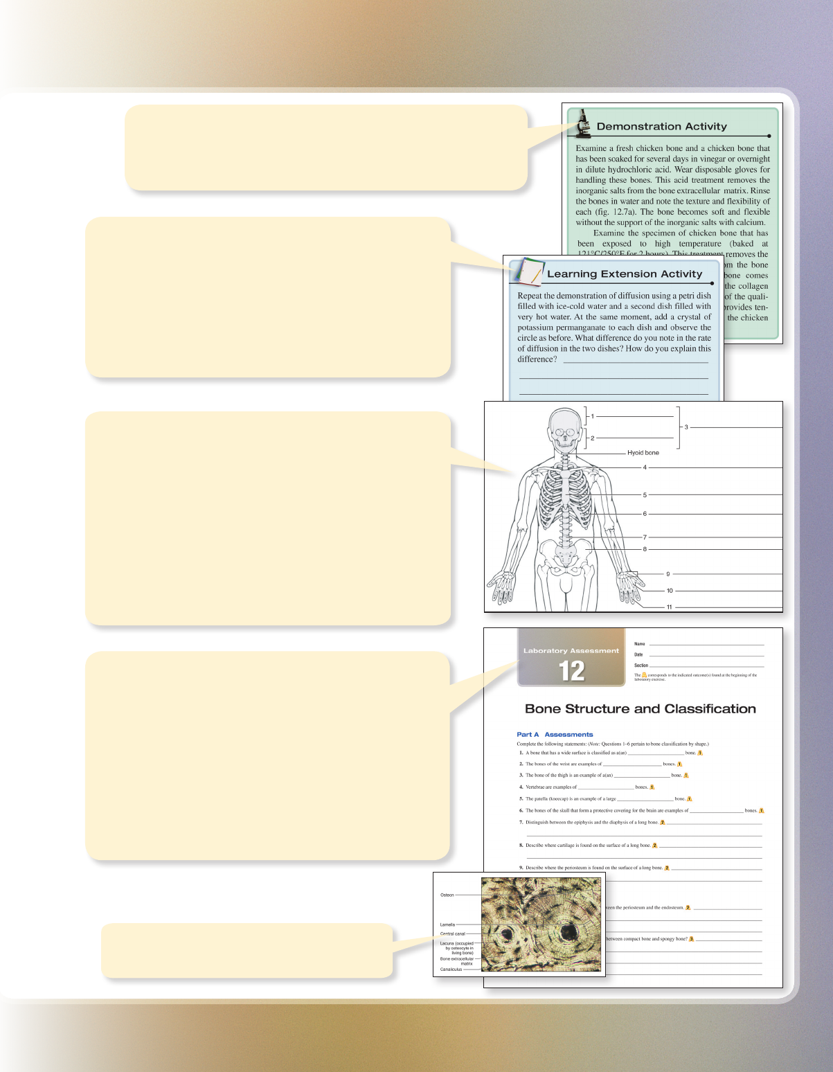
Learning Extension Activit ies Learning extension
activities also appear in separate boxes. They encourage
students to extend their laboratory experiences. Some of these
activities are open-ended in that they suggest the student plan
an investigation or experiment and carry it out after receiving
approval from the laboratory instructor. Some of the gures
are illustrated as line art or in grayscale. This will allow colored
pencils to be used as a visual learning activity to distinguish
various structures.
Illustrations Diagrams similar to those in a textbook
often are used as aids for reviewing subject matter. Other
illustrations provide visual instructions for performing steps
in procedures or are used to identify parts of instruments
or specimens. Micrographs are included to help students
identify microscopic structures or to evaluate student
understanding of tissues.
In some exercises, the gures include line drawings
suitable for students to color with colored pencils. This
activity may motivate students to observe the illustrations
more carefully and help them to locate the special features
represented in the gures.
Laboratory Assessments A laboratory assessment
form to be completed by the student immediately follows each
exercise. These assessments include various types of review
activities, spaces for sketches of microscopic objects, tables
for recording observations and experimental results, and
questions dealing with the analysis of such data.
As a result of these activities, students will develop a better
understanding of the structural and functional characteristics of
their bodies and will increase their skills in gathering
information by observation and experimentation. By completing
all of the assessments, students will be able to determine if
they were able to accomplish all of the learning outcomes.
Histology Histology photos placed within
the appropriate exercise.
Demonstration Activities Demonstration activities appear
in separate boxes. They describe specimens, specialized laboratory
equipment, or other materials of interest that an instructor may want
to display to enrich the student’s laboratory experience.
mar53064_FM.indd 11 9/14/11 8:44 AM

xii
Changes to This Edition
xii
Laboratory Exercise Topic Change
1 Laboratory Assessment Improved directions
2 Procedures A, B, and C
Structural lists
Laboratory Assessment
Added introductory material
Functions and descriptions added
Added content
3 Fig. 3.1 (pH values)
Procedure A
Improved depth
Added introductory material
4 Fig. 4.3 (microscope)
Laboratory Assessment
Added figure
Added content; improved accuracy in Part B
5 Fig. 5.1 (composite cell)
Introductory material
Ph.I.L.S. Lesson 2
Improved depth
Updated and expanded content
Clarity added
6 Procedures B, C, and D
Ph.I.L.S. Lesson 1
Added introductory material
Clarity added
7 Fig. 7.2 (interphase)
Fig. 7.5 (mitosis)
Fig. 7.6a (human chromosomes)
Introductory material
Added micrograph
Improved depth
New micrograph
Improved depth
8 Fig. 8.1a, b, d, g, and h (epithelial tissues)
Fig. 8.2 (sectional cuts)
Table 8.1 (epithelial tissues)
New micrographs
Added comparisons to body tube
Added table with descriptions, functions, and locations
9 Fig. 9.1b and h (connective tissues)
Table 9.1 (connective tissues)
Table 9.2 (connective tissues)
Introductory material
New micrographs
Added table with descriptions and functions
Improved design
Improved depth
10 Fig. 10.1a and c (muscle tissues)
Table 10.1 (muscle and nervous tissues)
New micrographs
Added table with descriptions, functions, and locations
11 Fig. 11.1 (skin layers)
Fig. 11.4b (skin structures)
Table 11.1 (epidermal layers)
Procedure
Added figure
New micrograph
Added table with locations and descriptions
Reworked
12 Fig. 12.1 (bone classification)
Fig. 12.4 (compact and spongy bone)
Procedure
Demonstration activity
Added figure
Added figure
Added introductory material
Rewritten information
13 Fig. 13.1a–b (skeleton)
Fig. 13.2a–h (bone features)
Introductory material
Redrawn
Added figure
Improved depth
14 Figs. 14.1 and 14.2 (skulls)
Fig. 14.7 (paranasal sinuses)
Table 14.1 (skull passageways)
Procedure
Redrawn
Added figure
Added table with locations and contents
Expanded depth
15 Fig. 15.5 (rib)
Procedures A and B
Structural lists
Added figure
Added introductory material
Added functions and descriptions
Global Changes
• Introductory materials expanded; introductory material precedes
most procedures.
• Pre-Lab questions expanded and placed in the laboratory
manual rather than online.
• BIOPAC exercises rewritten.
• Ph.I.L.S. laboratory simulations updated.
• Ph.I.L.S. 4.0 online included with lab manual.
• Structural lists have functions and descriptions added.
• Muscle tables added with origins, insertions, and actions.
• New design and sequence of items placed on the introductory
page of the laboratory exercise.
• Laboratory Reports changed to Laboratory Assessments.
• Matching assessments for the learning outcomes are all in the
Laboratory Assessments.
• Laboratory exercises contain fully labeled figures.
• Laboratory Assessments expanded and contain figures to label.
• All micrographs contain magnifications.
mar53064_FM.indd 12 9/14/11 8:44 AM

xiiixiii
Laboratory Exercise Topic Change
16 Fig. 16.2b (scapula)
Fig. 16.5 (hand bones)
Procedures A and B
Structural lists
Added figure
New figure
Added introductory material
Added functions and descriptions
17 Fig. 17.2a (hip bone)
Fig. 17.2b (hip bone)
Fig. 17.5 (foot bones)
Procedures A and B
Table 17.1 (male and female pelves)
Critical Thinking Activities
Revised figure
Added figure
New figure
Added introductory material
Added comparison table
Two added
18 Introductory material Improved depth
19 Fig. 19.2 (synovial joint)
Fig. 19.3b (cadaver knee)
Procedures A and B
Critical Thinking Activity
New figure
Added figure
Added introductory material
One added
20 Fig. 20.1 (neuromuscular junctions)
Fig. 20.3 (fascicle)
Fig. 20.5 (sarcomere)
Table 20.1 (muscle descriptions)
Structural list
Ph.I.L.S. Lesson 5
Added micrograph
Added micrograph
Added micrograph
Added table
Functions and descriptions added
Clarity added
21 BIOPAC Exercise (Electromyography) Rewritten
22 Tables 22.1, 22.2, 22.3, and 22.4 (head and neck
muscles)
Added tables with origins, insertions, and actions
23 Fig. 23.5a–b (forearm muscles)
Tables 23.1, 23.2, 23.3, and 23.4 (chest, shoulder,
and upper limb muscles)
Procedure
New figure
Added tables with origins, insertions, and actions
Reworked
24 Title and two procedures
Fig. 24.4a–c (pelvic floor muscles)
Tables 24.1, 24.2, and 24.3 (vertebral column,
abdominal wall, and pelvic floor muscles)
Improved topics, clarity, and depth
New figure
Added tables with origins, insertions, and actions
25 Fig. 25.5b (leg muscles)
Fig. 25.7 (leg muscles)
Tables 25.1, 25.2, and 25.3 (hip and lower limb
muscles)
Added figure
Added figure
Added tables with origins, insertions, and actions
26 Procedure
Laboratory Assessment Part C
Reworked sequence and clarity
Improved design and directions
27 Procedures A and B
Fig. 27.1 (structural neurons)
Fig. 27.6 (neuroglia)
Fig. 27.8 (Purkinje cell)
Fig. 27.9
Tables 27.1 and 27.2 (neurons and neuroglia)
New organization
Added figure
Added figure
Added micrograph
Added figure
Added tables with characteristics, locations, and functions
28 Title and three procedures
Fig. 28.3 (spinal cord)
Fig. 28.4 (spinal nerves)
Fig. 28.7 (meninges)
Fig. 28.8 (spinal cord)
Expanded topics, clarity, and depth
Expanded content
Added figure
Added figure
New micrograph
29 Fig. 29.1 (withdrawal reflex arc)
Fig. 29.2 (stretch reflex arc)
Laboratory Assessment Part A table
Expanded content
Added figure
Expanded components
30 Figs. 30.1 and 30.2 (ventricles of brain)
Fig. 30.7 (cerebellum and brainstem)
Tables 30.1, 30.2, and 30.3 (brain and cranial
nerves)
Procedure A
Added figures
Added figure
Added tables with descriptions and functions
Added introductory material
mar53064_FM.indd 13 9/14/11 8:44 AM

xiv
Laboratory Exercise Topic Change
31 BIOPAC (Electroencephalography) Rewritten
32 Fig. 32.3 (sheep brain)
Fig. 32.6 (sheep brain)
Redrawn figure
Added figure
33 Fig. 33.3 (two-point test)
Table 33.1 (skin receptors)
Added figure
Added table
34 Fig. 34.1 (smell receptors)
Fig. 34.4 (taste bud)
Revised figure
Revised orientation
35 Fig. 35.1 (lacrimal apparatus)
Fig. 35.6 (eye exam)
Fig. 35.12 (sectioned eye)
Table 35.1 (eye muscles)
Structural list
New figure
New figure
New micrograph
Added table with actions and nerves
Descriptions and functions added
36 Fig. 36.1 (refractive defects)
Procedure A
Added figure
Clarified directions
37 Structural list Descriptions and functions added
38 Laboratory exercise title Better reflects content of lab
39 Fig. 39.1 (major endocrine glands)
Fig. 39.6 (thyroid gland)
Fig. 39.12 (pancreas)
Ph.I.L.S. Lesson 19
New figure
New micrograph
New micrograph
Clarity added
40 Procedure A Added introductory material
41 Figs. 41.2 and 41.5 (blood cells)
Table 41.1 (blood components)
Introductory material
Added micrographs
Updated and expanded content
Improved depth
42 Procedure D (cholesterol test)
Ph.I.L.S. Lesson 34
Introductory material
Added procedure
Clarity added
Improved depth
43 Fig. 43.4 (blood test results)
Table 43.2 (blood typing reactions)
Added figure
Updated
44 Fig. 44.3 (sectioned heart)
Fig. 44.6 (blood circuits)
Structural list
Terminology
Added figure
Added figure
Added descriptions and functions
Updated
45 Fig. 45.1 (heart sound locations)
Fig. 45.4 (cardiac cycle)
Procedure B
Updated
Added figure
Added introductory material
46 BIOPAC (Electrocardiography) Rewritten
47 Fig. 47.1 (blood vessel wall structure)
Fig. 47.2 (artery and vein)
Fig. 47.6 (cerebral arterial circle)
Introductory material
Procedures A, C, and D
Added figure
New micrograph
Added figure
Improved depth
Added introductory material
48 Fig. 48.2 (taking pulse rate)
Fig. 48.5 (taking blood pressure)
Introductory material
Procedure B
Ph.I.L.S. Lesson 40
Added figure
Added figure
Improved depth
Added introductory material
Clarity added
49 Fig. 49.1 (fluid movements)
Fig. 49.2 (lymph drainage areas)
Fig. 49.6a (cadaver lymph node)
Fig. 49.6c (lymph node)
Fig. 49.7 (thymus)
Fig. 49.8 (spleen)
Introductory material
Added figure
Added figure
Added figure
New micrograph
New micrograph
New micrograph
Improved depth
Changes to This Edition
mar53064_FM.indd 14 9/14/11 8:44 AM

xv
Laboratory Exercise Topic Change
50 Fig. 50.1 (respiratory organs)
Fig. 50.5 (respiratory organs)
Fig. 50.6 (trachea wall)
Structural list
New figure
Added figure
New micrograph
Added descriptions and functions
51 Fig. 51.1 (respiratory muscles)
Fig. 51.2 (model for air movements)
Procedure A
Ph.I.L.S. Lesson 38
Added figure
Added figure
Added introductory material
Clarity added
52 BIOPAC (Spirometry) Rewritten
53 Figs. 53.1 and 53.2 (respiratory organs)
Fig. 53.3 (peripheral chemoreceptors)
Introductory material
New figures
Added figure
Updated and improved depth
54 Fig. 54.7 (stomach wall)
Structural lists
New micrograph
Added descriptions and functions
55 Fig. 55.1 (lock-and-key model)
Introductory material
Added figure
Improved depth
56 Laboratory exercise title
Fig. 56.2 (kidney section)
Fig. 56.6 (urethra of female and male)
Fig. 56.9 (urethra)
Procedure C
Structural lists
Reflects expanded content
Revised labels
Added figure
Added micrograph
Added introductory material and urethra
Added descriptions and functions
57 Procedures A and B Reflects new organization of contents
58 Fig. 58.1 (male reproductive system)
Fig. 58.2 (testis of cadaver)
Fig. 58.3 (testis)
Fig. 58.4 (seminiferous tubule)
Fig. 58.5 (epididymis)
Fig. 58.6 (ductus deferens)
Structural lists
Laboratory Assessments Part B
New figure
Added figure
Expanded labels
New figure
New figure
Added figure
Added descriptions and functions
New content arrangement
59 Fig. 59.3 (female cadaver organs)
Fig. 59.8 (uterine tube)
Fig. 59.9 (uterine wall)
Structural lists
Laboratory Assessments Part B
Added figure
New micrograph
New micrograph
Added descriptions and functions
New content arrangement
60 Structural list Added descriptions and functions
61 Introductory material Improved depth
mar53064_FM.indd 15 9/14/11 8:44 AM
